mail_outline sales@mediastorehouse.com
Choose a picture from our collection for your Wall Art and Photo Gifts
438 Items
Pleurosigma angulatum enlarged under a microscope. Date of Photograph:04/1893
Microscopic enlargement of a micro-organism on a slide. Date of Photograph:1870-1890 ca
Microscopes Koriska, School of Science in the Real Commercial Technical Institute F. Besta in Treviso Treviso. Date of Photograph:1950-1960
Microscope Koriska, School of Science or Merceology Museum in the Real Commercial Technical Institute F. Besta in Treviso Treviso. Date of Photograph:1950-1960
Laboratory of Merceology at the Technical Institute of Bolzano; on the counter some microscopes and a measuring instrument for flour moisture measurement Bolzano. Date of Photograph:1930-1940
A radiologist takes a radiograph of a patient with the Microscope 500. Instrument, discovered in America, which is much more protective to radiation. America. Date of Photograph:1950 - 1960
Biology. Microscopic enlargment of puccinia graminis microorganisms, wheat parasites. Date of Photograph:1870-1890 ca
Microscopic study of a cell of nummulite on a slide. Date of Photograph:1870-1890 ca
Biology. Microscopic enlargement of a micro organism on a slide. Date of Photograph:1870-1890 ca
Planoarcina Ureae enlarged under a microscope. Date of Photograph:1914 ca
Microscopic enlargement of Proteus Vulgaris. Date of Photograph:1914 ca
Microscopic enlargement of the cholera vibrio. Date of Photograph:1872-1915 ca
A bacillus enlarged under a microscope. Date of Photograph:1890-1905 ca
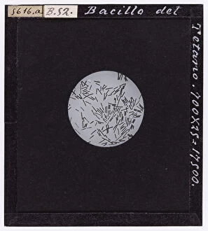
Microscopic enlargement of the Tetanus bacillus. Date of Photograph:1872-1915 ca
Sporiferous tetanus bacillus enlarged under a microscope. Date of Photograph:1872-1915 ca
Streptococcus: bacteria enlarged under a microscope. Date of Photograph:1888-1915 ca
Female Anopheles simplex, magnified under the microscope. Date of Photograph:1890-1914 ca
Male Anopheles simplex, magnified under the microscope. Date of Photograph:1890-1914 ca
Female Anopheles maculiformis, magnified under the microscope. Date of Photograph:1890-1914 ca
Microscopic enlargement of Achorion Schoenleinii, parasite fungus of man. Date of Photograph:1897-1914 ca
Fabulous ringworm fungus on day 16 magnified under the microscope. Date of Photograph:1914 ca
Campanularia Dichotoma (Campanularie) Hydromedusa, enlarged under the microscope. Date of Photograph:1914
Culture of Pseudomonas Porrettana in colonies, daisy-shaped magnified under the microscope. Date of Photograph:01/1905
Gonothyraea Loveni (Polyp-medusae) magnified under the microscope. Date of Photograph:1897
Phosphorescent bacillus, enlarged under a microscope, photographed with the same light after an hour of air exposure. Date of Photograph:1890-1905 ca
Pseudomonas Porrettana enlarged under a microscope. Date of Photograph:03/1905
Phosphorescent bacillus with cillia found in an omelette. Enlarged under a microscope. Date of Photograph:02/1903
Pyogenus foetidus bacillus enlarged under a microscope. Date of Photograph:01/1890
Microscopic enlargment of bacillus of Anthrax nested in a kidney glomerule of a Guinea pig. Date of Photograph:1900-1915 ca
Diptheric bacillus, enlarged under a microscope. Date of Photograph:1872-1915 ca
Bacillus Chauvaei, enlarged under a microscope. Date of Photograph:1872-1915 ca
Sputum with tubercular bacillus, enlarged under a microscope. Date of Photograph:1888-1915 ca
Culture of tubercular bacillus, enlarged under a microscope. Date of Photograph:1888
Gonococco: bacteria enlarged under a microscope. Date of Photograph:1888-1915 ca
Morphology of bacteria, various aspects in reproduction, microscopic enlargement, plate 5, in W. Migula "System der Bakterien "Handbuch der Morphologie
Morphology of bacteria, polar granules, lacunae, capsules and pseudo-capsules, microscopic enlargement, plate 47, in W. Migula System der Bakterien "Handbuch der Morphologie
Microscopic enlargment of Saccharomyces Equi. Date of Photograph:1902-1913 ca
Saccharomyces apiculata: microscopic, unicellular fungus, of the Ascomycetes, enlarged under a microscope. Date of Photograph:04/1913
Microscopic enlargment of Saccharomyces Cerevisiae. Date of Photograph:04/1913
Crenothrix polyspora from a Florentine aqueduct magnified under the microscope. Date of Photograph:1913
The bacterium Beggiatoa alba magnified under the microscope. Date of Photograph:04/1913
Gelatinous layer on the surface of the waters of the Donzelle spring enlarged under the microscope, Porretta. Date of Photograph:1905
Phragmidium asperum fungus, belonging to the Uredineae, enlarged under a microscope. Date of Photograph:04/1914
Puccinia graminis: fungus belonging to the Uredineae, enlarged under a microscope. Date of Photograph:1914
Fungus belonging to the Uredineae, called Uromyces Ficaria, enlarged under a microscope. Date of Photograph:12/1902
Uredineae: parasite fungus that invades the Graminaceae producing the disease called rust. Enlarged under a microscope. Date of Photograph:12/1902
Melampsora salicina fungus, belonging to the Uredineae class, enlarged under a microscope. Date of Photograph:12/1902
Puccinia arundinacea, belonging to the Hyphomycetes, enlarged under a microscope. Date of Photograph:1898