mail_outline sales@mediastorehouse.com
Malaria plasmodium enlarged under a microscope. Summer-Fall fever. Semi-lunar shapes. Date of Photograph:1872-1915 ca
Roman mosaic from Porto Torres, now in the G.A. Sanna National Museum in Sassari Sassari National Museum Mosaic Roman Art, Europe, Ancient Civilization. Date of Photograph:1938 ca
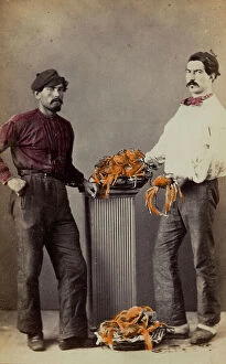
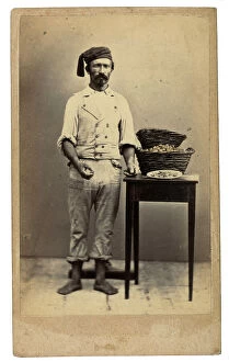
Venetian costumes and crafts: Peddlers of edible crabs Venice. Date of Photograph:1890 ca
Still life with fish, birds and vegetables Great Britain. Date of Photograph:1865 ca
Skeleton of an insect Great Britain. Date of Photograph:1865 ca
Winner of the Festa del Grillo (Cricket Festival) Florence. Date of Photograph:1930 ca
Postcard, portrait of a little girl with butterfly wings and flower, "Album para Tarjetas postales" France. Date of Photograph:1910 ca
Postcard depicting a woman in a forest with a butterfly, "Album para Tarjetas postales" Europe. Date of Photograph:1910 ca
An oyster (ostrea edulis) in a rocky sea depth. Europe. Date of Photograph:1960-1965 ca
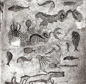
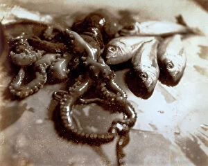
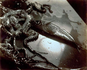
Still life with fish and polyps. Date of Photograph:1934
Still life with polyps and fish. Date of Photograph:1934
Pictorialist still life with fish and polyps. Date of Photograph:1934
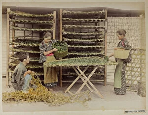
Two young Japanese women in traditional costume during the processing of silkworms Japan. Date of Photograph:1885-1895
Venetian costumes and masks: peddler of mussels and oysters Venice. Date of Photograph:1860 ca
Rearing of silkworms for the textile industry, Japan Japan. Date of Photograph:1890-1899
Album "Von Herr August Egli in Jokohama - Juni 1895": two young Japanese women in traditional costume settle silkworms useful to the textile industry Japan Yokohama
Two butterflies on the leaf of a plant. Europe. Date of Photograph:1955 - 1960
Fragment of an Egyptian bas-relief from the Temple of Usertesen I, in the British Museum in London Great Britain London, British Museum Low Relief, Basrelief Egypt - Egyptian Art, Africa
Calliphora Vomitoria (Diptera. Muscidae), from A. Celli, "Manuale dell'igienista, ad uso di ufficiali sanitari, medici circondariali e provinciali, ingegneri, chimici e veterinari igienisti
Sarcophaga carnaria (Diptera)(Muscidae) from A. Celli, "Manuale dell'igienista, ad uso di ufficiali sanitari, medici circondariali e provinciali, ingegneri, chimici e veterinari igienisti
Female Anopheles simplex, magnified under the microscope. Date of Photograph:1890-1914 ca
Male Anopheles simplex, magnified under the microscope. Date of Photograph:1890-1914 ca
Female Anopheles maculiformis, magnified under the microscope. Date of Photograph:1890-1914 ca
Hydatina scrita and Hematococcus nivalis protozoa enlarged under a microscope. Date of Photograph:1913
Hydatina scrita and Hematacoccus nivalis micro-organisms, enlarged under a microscope. Date of Photograph:04/1913
Ciliate protozoa, Paramecium putrinum, enlarged under a microscope. Date of Photograph:04/1914
Carchesium polypinum: ciliate protozoa enlarged under a microscope. Date of Photograph:1897
Negri bodies present in the cell of the of the Horn of Ammon of a rabid cow, enlarged under a microscope. Date of Photograph:1900-1915 ca
Protozoa of babesia canis in bone marrow: blood parasite in mammals, enlarged under a microscope (Haemosproidia). Date of Photograph:1872-1915 ca
Babesia canis in bone marrow, enlarged under a microscope. Date of Photograph:1872-1915 ca
Plasmodium of the Colobus Guereza monkey, enlarged under a microscope. Date of Photograph:1872-1915 ca
Microscopic enlargement of the parasite of the Tertian fever. Date of Photograph:1900-1915 ca
Microscopic enlargment of the malaria cists in the stomach of Anopheles, the malaria mosquito. Date of Photograph:1900-1915 ca
Miocroscopic enlargement of Sporozoi, parasite protozoans. Date of Photograph:1888-1915 ca
Clamidococcus nivalis (Alga) enlarged under a microscope. The protozoa fell with the rain in October 1900, in Florence. Date of Photograph:12/1902
Haematococcus pluviatis: Phitomonadino, enlarged under a microscope. Date of Photograph:1888
Section of the stomach of an Anopheles, Amfionte (Zygote); fertilized macrospore in this image, enlarged under a microscope. Date of Photograph:1888
Types of Tripanosoma Abissinicum, Theileri, and Piroplasma ligem, belonging to the Bovis species, enlarged under a microscope. Date of Photograph:1872-1915 ca
Protozoa flagellata: Tripanosoma abissinicum found in the ox, enlarged under a microscope. Date of Photograph:1872-1915 ca
Chrysomyia macellaria (Diptera. Muscidae) from A. Celli, "Manuale dell'igienista, ad uso di ufficiali sanitari, medici circondariali e provinciali, ingegneri, chimici e veterinari igienisti
Female Anopheles punctatus, magnified under the microscope. Date of Photograph:1890-1914 ca
Spirochaeta pallida bacterium: unicellular micro-organism with a stretched spiral shape, enlarged under a microscope. Date of Photograph:1900-1915 ca
Paramoecion putrinum: ciliate protozoa enlarged under a microscope. Date of Photograph:1897
Bigeminum babesia in oxen: protozoa enlarged under a microscope(Haemosproidia). Date of Photograph:1872-1915 ca
Tropical babesia in oxen: illness owed to parasite protozoa (pyroplasma) in the blood of mammals, transmitted by ticks (Haemosproidia). Enlarged under a microscope. Date of Photograph:1872-1915 ca
Pigeon blood with Datulewski Plasmodium, enlarged under a microscope. Date of Photograph:1900-1915 ca
Ceratium tripos: protozoa belonging to the Flagellati, enlarged under a microscope. Date of Photograph:04/1914
Hematococcus nivalis, belonging to the Clamido-monadine family, enlarged under a microscope. Date of Photograph:04/1913